Fruit under microscope reveals a world of intricate structures and mesmerizing patterns that few people ever get to see. By examining fruits at a microscopic level, we gain a deeper understanding of their composition, structure, and the remarkable processes that make them vital to life on Earth. Whether you're a botanist, food scientist, or simply a curious individual, this microscopic exploration offers a unique perspective on something we often take for granted.
The study of fruits under a microscope is more than just a scientific curiosity. It bridges the gap between biology, agriculture, and nutrition, providing valuable insights into the mechanisms that govern plant reproduction and fruit development. In this article, we will delve into the fascinating world of fruits at a microscopic scale, exploring their cellular structures and the roles they play in ecosystems.
Whether you're looking to deepen your knowledge or simply satisfy your curiosity, this article will guide you through the wonders of fruit anatomy under magnification. From the cellular walls of apples to the intricate seed patterns of oranges, every fruit tells a story under the lens of a microscope.
Read also:Liam Flockhart The Rising Star In The Entertainment Industry
Table of Contents
- Introduction to Microscopy in Studying Fruits
- Fruit Cell Structure: The Building Blocks
- Microscopic Examination of Common Fruits
- Fruit Under Microscope: Apple
- Fruit Under Microscope: Orange
- Fruit Under Microscope: Grape
- Tools and Techniques for Fruit Microscopy
- Scientific Applications of Fruit Microscopy
- The Role of Microscopy in Understanding Fruit Nutrition
- Future Directions in Fruit Microscopy Research
Introduction to Microscopy in Studying Fruits
Microscopy has long been an essential tool in the study of plant biology, offering scientists and enthusiasts alike a window into the microscopic world of fruits. By magnifying fruit tissues, researchers can observe cellular structures, identify pathogens, and study developmental processes. The use of microscopes in fruit research has paved the way for advancements in agriculture, food science, and environmental studies.
Modern microscopes, ranging from light microscopes to electron microscopes, provide varying levels of magnification and detail. Each type of microscope reveals different aspects of fruit anatomy, allowing for comprehensive analysis. For example, electron microscopes can capture ultra-high-resolution images of fruit cell walls, while light microscopes are ideal for observing stained tissue samples.
Why Study Fruits Under a Microscope?
Studying fruits under a microscope offers numerous benefits:
- Insight into cellular composition and structure
- Identification of diseases and pests affecting fruit crops
- Understanding the mechanisms of nutrient storage and transport
- Development of new techniques for improving fruit quality and yield
Fruit Cell Structure: The Building Blocks
Fruits are composed of various types of cells, each with specific functions that contribute to the overall structure and function of the fruit. The primary components include epidermal cells, mesocarp cells, and endocarp cells, among others. Each cell type plays a crucial role in the fruit's development and nutritional value.
Epidermal cells form the outer layer of the fruit, providing protection against environmental stressors such as UV radiation and pathogens. These cells often contain cutin or wax, which helps to retain moisture and prevent dehydration.
Key Features of Fruit Cells
- Cell walls rich in cellulose and pectin
- Vacuoles storing water, sugars, and pigments
- Chloroplasts in green fruits for photosynthesis
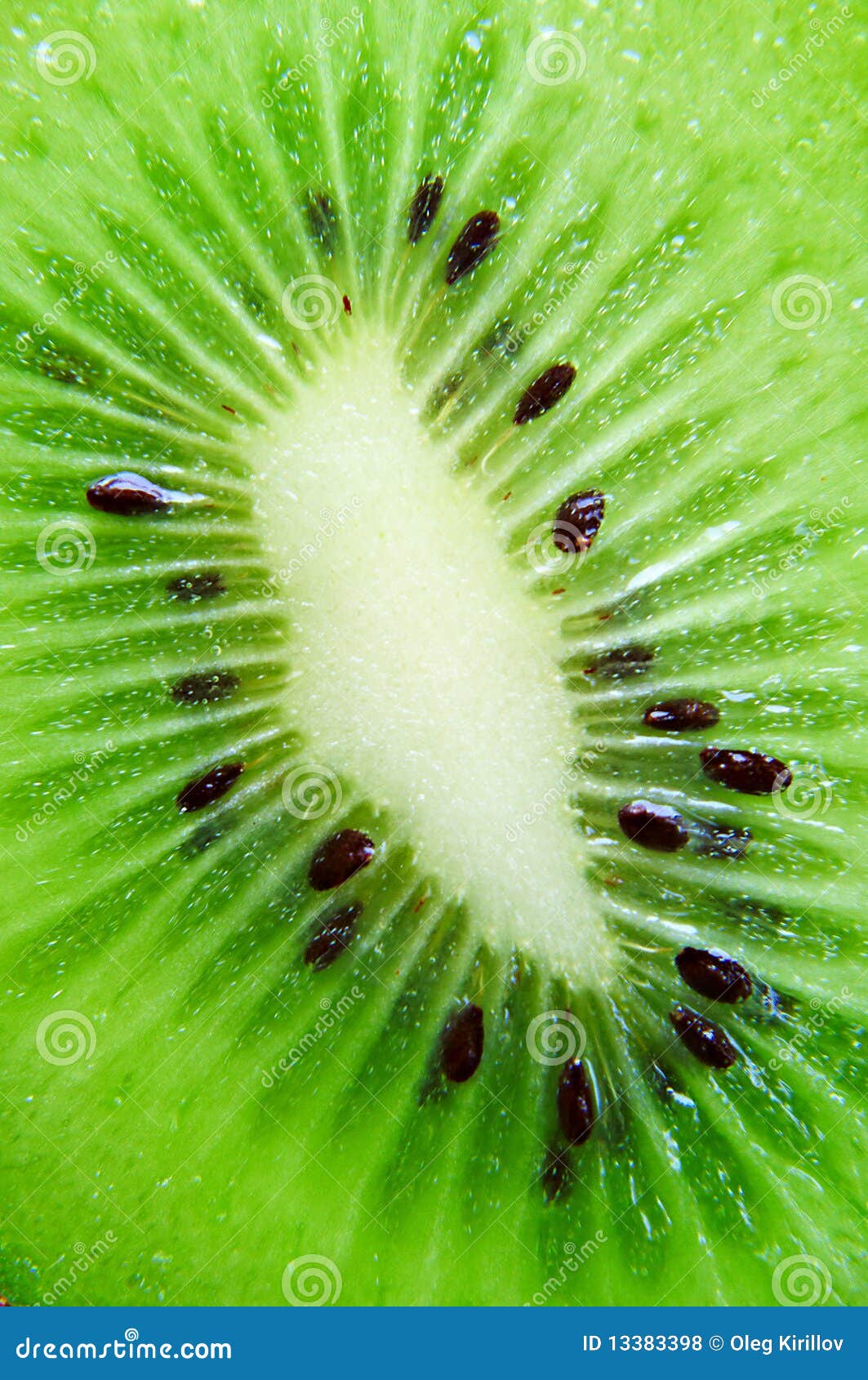
Microscopic Examination of Common Fruits
Examining common fruits under a microscope reveals a wealth of information about their structure and function. From the juicy pulp of citrus fruits to the fibrous flesh of apples, each fruit has its own unique characteristics that can be observed at a microscopic level.
Read also:Amber Heard On Top Gear A Comprehensive Look At Her Appearance And Impact
Fruit Under Microscope: Apple
Apples are a popular fruit known for their crisp texture and sweet flavor. Under a microscope, apple cells exhibit a distinct arrangement of parenchyma tissue, which gives the fruit its characteristic texture. The cell walls are thick and rich in pectin, contributing to the fruit's firmness.
Key observations:
- Parenchyma cells with large vacuoles
- Thickened cell walls containing pectin
- Starch grains visible in immature apples
Fruit Under Microscope: Orange
Oranges are renowned for their high vitamin C content and juicy segments. Microscopic examination of orange tissue reveals a network of juice sacs, or vesicles, that store water and nutrients. These sacs are surrounded by thin-walled cells that facilitate the transport of sugars and acids.
Key observations:
- Juice sacs filled with water and nutrients
- Thin-walled cells for efficient transport
- Pigmented cells contributing to the fruit's color
Fruit Under Microscope: Grape
Grapes are another fruit that reveals fascinating details under a microscope. The skin of a grape is composed of thick epidermal cells that protect the delicate flesh inside. The flesh itself consists of soft parenchyma cells filled with sugary sap.
Key observations:
- Thick epidermal cells forming the skin
- Soft parenchyma cells in the flesh
- Pigmented cells responsible for color variation
Tools and Techniques for Fruit Microscopy
Effective fruit microscopy requires the right tools and techniques. From basic light microscopes to advanced electron microscopes, each instrument offers unique capabilities for studying fruit anatomy. Staining techniques, such as using iodine or safranin, enhance visibility of cellular structures, making it easier to identify key features.
Key tools:
- Light microscope for general observations
- Electron microscope for high-resolution imaging
- Staining solutions for enhancing contrast
Scientific Applications of Fruit Microscopy
The study of fruits under a microscope has far-reaching implications in various fields. In agriculture, microscopy aids in the identification of diseases and pests, helping farmers to implement targeted pest management strategies. In food science, it provides insights into the processes of ripening and spoilage, leading to improved storage and preservation techniques.
Environmental scientists also benefit from fruit microscopy, as it helps them understand the role of fruits in ecosystems and their interactions with pollinators and seed dispersers.
The Role of Microscopy in Understanding Fruit Nutrition
Fruit microscopy plays a critical role in nutritional research, allowing scientists to study the distribution and composition of nutrients within fruits. By examining the cellular structures responsible for storing vitamins, minerals, and antioxidants, researchers can develop strategies to enhance the nutritional value of fruits.
Data from studies suggest that certain fruits, such as berries and citrus fruits, are particularly rich in bioactive compounds that promote health and well-being. Microscopy helps to identify these compounds and understand their mechanisms of action.
Future Directions in Fruit Microscopy Research
As technology continues to advance, the field of fruit microscopy is poised for exciting developments. Innovations in imaging techniques, such as confocal microscopy and super-resolution microscopy, promise to reveal even more intricate details about fruit anatomy. These advancements will enable researchers to explore new frontiers in plant biology, agriculture, and nutrition.
Future research may also focus on the application of artificial intelligence and machine learning in analyzing microscopic images, streamlining the process of data collection and interpretation.
Conclusion
The world of fruits under a microscope is a captivating one, filled with intricate structures and fascinating processes. From the cellular composition of apples to the juice sacs of oranges, every fruit tells a unique story when viewed at a microscopic level. This exploration not only deepens our understanding of fruit anatomy but also has practical applications in agriculture, food science, and environmental studies.
We invite you to join the conversation by leaving a comment below or sharing this article with others who may find it interesting. For further reading, explore our other articles on plant biology and microscopy. Together, let's continue to uncover the wonders of nature's tiny wonders!